Summary
The AED designed and constructed two corers specifically for collecting intact (undisturbed) sediment cores for CT analysis. Since any high-density materials such as metals can interfere with CT X-ray imaging, all core tube parts were constructed from PVC and nylon plastic.
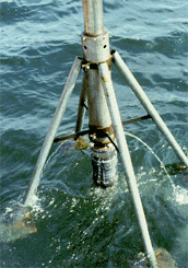
Corer used for water depths greater than 3 meters. Photo 1, shows the corer used in water depths >3 meters. |
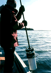
Corer used for shallow water Photo 2, is the hand held corer used in shallow water. The sediments are routinely collected in into 15cm diameter x 30 to 45 cm long PVC tubes. In each corer the sediment is held securely within the tubes after collection with a flapper valve. |
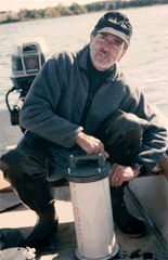
Corer used for soft bottom sediments Photo 3, illustrates a core tube being assembled after sampling sediment. |
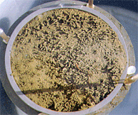
Intact sediment taken, without physical disruption, by one of our corers. Photo 4, shows an example of an intact sediment core taken with one of our corers and illustrates that the coring operation causes very little physical disruption at the sediment-water interface (SWI). The cores, with the tops removed, can be held in flowing seawater until they are scanned; however, they are usually CT scanned at our local hospital, in the evening of the same day of collection. |
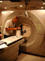
Picker 2000 CT medical scanner Photo 5, shows a sediment core being aligned prior to scanning in a Picker 2000 CT. |
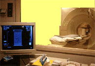
A computer interface from which scanned images are transferred to computer tape. |
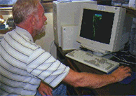
The computer interface used at the EPA laboratory to which image data was transferred |
CT Home | Introduction | Methods | 3-D CT Movies | Summary | Glossary | Literature Cited | Contacts
![]()
![[logo] US EPA](../gif/logo_epaseal.gif)