Introduction
Computer Aided Tomography (CT) imaging, usually used by physicians to diagnose medical problems in human patients, is being applied by scientists at the Environmental Protection Agency (EPA-ORD-NHEERL-AED) in Narragansett, RI, USA to evaluate the ecological state of marine sediment communities. This is the first time ecologists have used this powerful technology to study environmental pollution problems (Perez et al. 1999). Traditionally ecological state is determined by collecting, identifying and counting sediment macrofauna; however, this procedure requires specialized training and is labor intensive and time consuming. CT offers a rapid, cost-effective additional measure to this traditional method by quantifying the burrows and tubes that these organisms create.
Investigators have attempted to detect and measure ecological changes due to natural and anthropogenic stresses. Generally, communities under stress lose species. Pearson and Rosenberg (1978) implied (refer to Figure 1) that in marine sediment communities, a direct relationship exists between changes in species, and the degree of tube and tunnel formation. They assumed that these changes were due mainly to the level of organic loading and portrayed the replacement of large, deeper tubes by smaller, more abundant surface tubes as organic load increases.
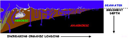
As the organic load gradient increases, the larger and deeper burrowing macro fauna in aerobic sediments are replaced by smaller species that live in close proximity to the sediment water interface. Figure 1: Along a gradient of increasing organic loading, the larger and deeper burrowing macrofauna in sediments are replaced by smaller species that live in close proximity to the sediment-water interface (SWI) . Scientists have used various techniques to investigate the relationship between species and the physical structures they create. Only recently has CT been applied to environmental and biological research. Because it can distinguish differences in object density, as measured by X-ray penetration, Warner et al. (1989) and Daniel et al. (1997) were able to quantify the vertical distribution of macrofaunal tubes and tunnels in terrestrial cores. In sediments from aquatic systems, Fu et al. (1994) and Magwood and Ekdale (1994) made qualitative examinations of biologically derived internal structures. Orsi et al. (1994) published the first three-dimensional (3D) image of marine sediment analyzed by CT. Recently, Michaud et al. (2003) used CT to follow temporal changes of benthic communities in an unstable sedimentary environment. |
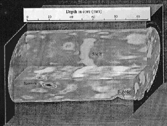
This image depicts the first 3-dimensional image of a marine sediment created by Orsi, et al., 1994 Figure 2. The graphic to the right depicts the first 3D image of a CT analyzed marine sediment that was created by Orsi et al. (1994). The U.S.E.P.A.'s Atlantic Ecology Division (AED) developed CT procedures (Perez et al.1999) to quantify both the percentage of macrofaunal tube and tunnel area (PTTA) in "soft" bottom marine sediments, as well as, sediment X-ray attenuation (SXA) . |
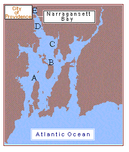
Map of Narragansett Bay with sampling sites where variables PTTA and SXA were measured Figure 3. Our first study (Perez et al. 1999) examined PTTA and SXA along a 31-km pollution gradient (Pruell and Quinn 1985) in Narragansett Bay, RI, USA. Sediment pollutants were highest at Station E and decreased to background levels at Station A. This embayment conformed to the Pearson and Rosenberg (1978) model and had increasing numbers of species related to decreasing degree of sediment pollution (Perez et al. 1990). |
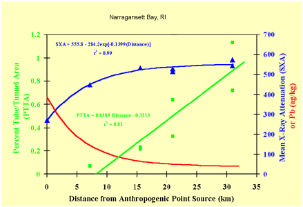
This graph illustrates the quantitative application of CT data to the determination of a marine pollution gradient Figure 4. Illustrates the relationship of quantitative CT measurements and sediment pollution concentrations along a marine pollution gradient (Narragansett Bay, RI, USA). The red curve represents the decrease in concentration of lead in sediment with distance from the polluted northern reach of Narragansett Bay. Many other anthropogenic contaminant metals and organics follow this same pattern. The green curve represents the increase in the percentage of macrofaunal tube and tunnel area (PTTA) and the blue curve represents the increase in the sediment X-ray attenuation (SXA) with distance from the polluted northern reach of Narragansett Bay. |
CT Home | Introduction | Methods | 3-D CT Movies | Summary | Glossary | Literature Cited | Contacts
![]()