Extramural Research
Presentation Abstract
Grantee Research Project Results
Title of Talk:
A Bioengineering Approach to Nanoparticle-Based Environmental Remediation
Abstract of Talk:
Research has investigated the potential use of ferritin, and ferritin-derived compounds, as templates to synthesize nanomaterials and to use these materials as substrates for environmental degradation processes. Specifically, recent research has developed a synthetic route for the assembly of supported nano metal oxide particles by assembling them in the biological protein ferritin and then removing the protein shell. This methodology has been further exploited to allow us to produce nanometallic particles of homogeneous size. Also, recently investigated has been the fundamental chemistry of ferritin-derived systems and to investigate their activity for the remediation of toxic metals in the presence and absence of light. Selected recent results are now shown.
Horse spleen Ferritin (HS-Fn) as a precursor to supported metal oxide and metallic nanoparticles (1):
Figure 1 shows a Tapping mode™ AFM image of ferrihydrite nanoparticles prepared by UV-ozone treatment of 2500 Fe loaded ferritin dispersed on a SiO2 substrate. The accompanying cross-section shows the full range of height values to be 6.5 nm, with a root-mean-square (RMS) roughness (standard deviation of the height about the average value) of 1.46 nm. The peak-to-valley height differences for the large features in the cross-section are in the 4-6 nm range. The lateral full widths at half maximum (FWHM) of the individual particles are several times larger than the heights due to tip convolution effects.
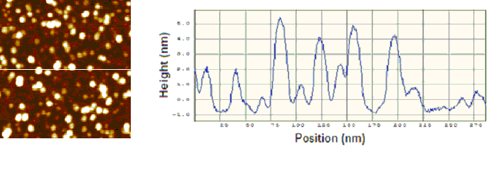
Figure 1: AFM Tapping mode™ micrographs (top view)
of FeOOH nanoparticles prepared by uv-ozone treatment of 2500 Fe loaded
ferritin for 60 mins at 100oC under oxygen (<5psi); section analysis
Reduction of the ferrihydrite nanoparticles to the Fe metal was carried out in a reaction cell coupled to the ultra-high vacuum (UHV) chamber. The sample was then transferred directly into the UHV chamber where x-ray photoelectron spectroscopy (XPS) was carried out. The AFM image of these particular particles (Fig. 2) shows a similar morphology to the particles prior to reduction. Again, the peak-to-valley height differences for the large features in the cross-section are in the 4-6 nm range, and in this case the RMS roughness is 1.47.
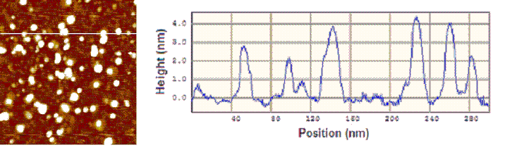
Figure 2: AFM Tapping mode™ micrographs (top view)
of iron nanoparticles prepared by heating 2500 Fe loaded FeOOH nanoparticles
in a reducing environment; section analysis
Formation of reactive Cu nanoparticles encapsulated within the protein
cage of ferritin (2):
The photolysis of Cu(II) in the presence of iron oxide-mineralized ferritin
and a sacrificial reductant such as citrate or tartrate resulted in the
formation of a wine-red color after an hour. Control reactions photolyzed
in the absence of Cu(II), tartrate/citrate, or iron oxide mineralized
ferritin, did not change color over the same period. No reaction was observed
when solutions were left in the dark or in the presence of the unmineralized
(apo) ferritin. In addition, Fe(II) alone was not observed to spontaneously
reduce Cu(II) in deaerated solutions. Examination of the photolysis products
by transmission electron microscopy (TEM) revealed electron-dense spherical
particles (see Figure 3), with the Cu(II):ferritin ratio serving as the
major determinant of particle size. Histograms of particle sizes were
fit to Gaussian distributions. Higher Cu(II):ferritin ratios led to larger
particle sizes, with loadings of 250, 500, 1000, and 2000 leading to average
particle diameters of 4.5 ± 0.8, 9.7 ± 4.2, 12.7 ±
3.6, and 31.4 ± 10.1 nm, respectively. The reactivity of these
nano-Cu particles will be investigated with regard to environmental remediation
reactions.
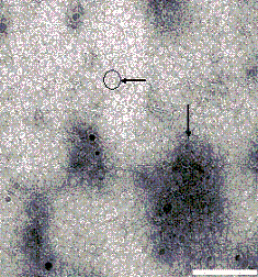
Figure 3: Metallic Cu particles grown within ferritin.
Formation of Fe2O3 and TiO2 nanoparticles within the protein cage of ferritin:
Mineralization of mammalian ferritin using high oxidation state transition metal ions as starting materials can be achieved using a photoreduction process. We have successfully mineralized the Fn cages with Fe-oxhydroxide and Ti-oxhydroxide nanoparticles, in a spatially selective manner, and encapsulated the resultant nanoparticles within the protein cages. The composite materials, synthesized in this way, have particle sizes that are highly monodisperse. This represent a new synthetic approach to the formation of protein encapsulated metal oxhydroxide materials in the nanoscale size regime.
Photolysis of a solution of Fe(III) or Ti(IV) citrate in the presence of the apo-ferritin cage results in the formation of small amounts of Fe(II) and Ti(III) respectively. In the presence of oxygen these reoxidize and hydrolyze to form nanoparticles within the protein cage. The Fe reaction could be monitored spectroscopically, by the development of the ligand-to-metal charge transfer absorbance characteristic of Fe(III)-oxo polymers. When the reaction was performed in the absence of the protein cages the development of the color was clearly associated with the formation of a rust colored precipitate. When Ti(IV)citrate was used as a starting material there was no change in color during the photolysis reaction, but in the presence of the protein cages the reaction solution remained homogeneous while in control reactions, in the absence of the protein cage, a white precipitate formed as the photolysis proceeded. If the photolysis was performed under inert atmosphere, the characteristic purple color of d1 Ti(III) species could be observed.
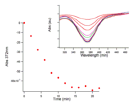
Figure 4: The plot shows the loss of Cr(VI) (reflected
in the loss of absorbance at 372 nm) from solution as the Ti-oxyhydroxide
catalyzes its reduction to Cr(III) in the presence of light.
The Ti-oxyhydroxide material was tested for its ability to catalyze the photo-reduction of CrO42- under Xe-arc lamp illumination. This material exhibited a catalytic behavior similar to that observed previously for Fe2O3-Ferritin (3).
References
(1) Iron and Cobalt Nanoparticles Engineered by the Reduction of Ferritin Encapsulated Metal Oxide Precursors, Hazel-Ann Hosein, Daniel R. Strongin, and Trevor Douglas, in review, Langmuir (2004).
(2) Photocatalytic Synthesis of Copper Colloids from Cu(II) by the Ferrihydrite Core of Ferritin, Daniel Ensign, Mark Young, and Trevor Douglas, submitted to Inorganic Chemistry 43(11) (2004) 3441-3446.
(3) Photochemical reactivity of ferritin for Cr(VI) reduction," Kim I., Hosein H., Strongin D.R., and Douglas T. Chemistry of Materials 14 (2002) 4874-4879